
MICCAI 2015, the 18th International Conference on Medical Image Computing and Computer Assisted Intervention, will be held from October 5th to 9th, 2015 in Munich, Germany.
MICCAI attracts annually world leading scientists, engineers and clinicians from a wide range of disciplines associated with medical imaging and computer assisted surgery.
The conference series includes three days of oral presentations and poster sessions. It also includes workshops, tutorials and challenges just before and after the conference.
We have a chance to be a part of this event because CancerCenter team starts in 2 challenges about „Imaging & Digital Pathology”.
Challenges which we joined:
– Challenge 1: Segmentation of Nuclei in Digital Pathology Images.
– Challenge 2: Combined Imaging and Digital Pathology Primary Tumor Classification.
Top three winners of each challenge will give brief presentations of their algorithms during the afternoon challenge sessions.
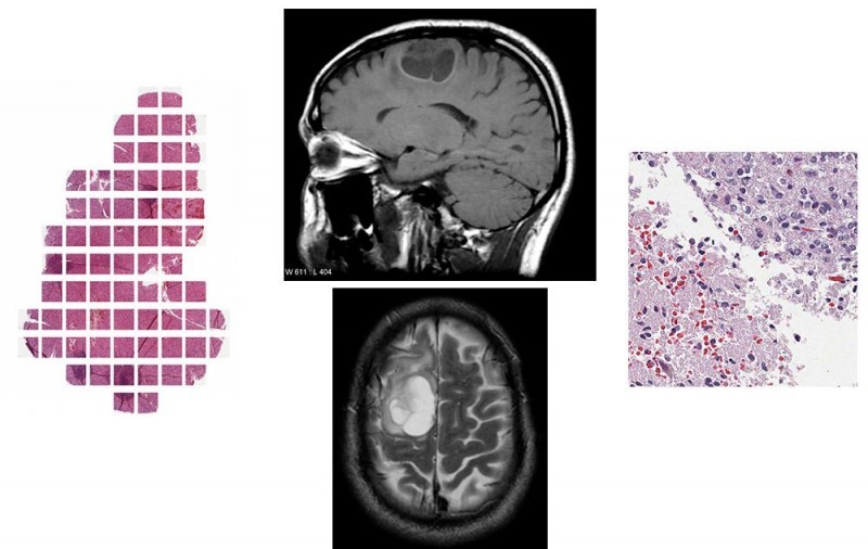
Informations about glioma:
A glioma is a type of brain tumor that grows from glial cells. Glial cells support nerve cells with energy and nutrients and help maintain the blood-brain barrier. Glioma is an umbrella term used to describe the different types of gliomas: astrocytoma, oligodendroglioma, and glioblastoma. Gliomas vary in their aggressiveness, or malignancy. Some are slow-growing and are likely curable. Others are fast-growing, invasive, difficult to treat, and are likely to recur.
Types of Glioma:
I – Pilocytic astrocytoma
II – Low-grade glioma
III – Malignant glioma Glioblastoma multiforme (GBM) – GBM is the most aggressive and most common primary brain tumor.
Image analysis for computer-aided medical diagnosis is a rapidly growing discipline of scientific research.
Many medical tasks can be automated automated, which
not only is economically beneficial but above all allows doctors to devote more time to patients. That is why is so important and excepted progress in digital image diagnostic.