PET Analysis

What is a PET/CT scan?
PET (Positron Emission Tomography) and CT (Computed Tomography) are both standard imaging tools that physicians use to pinpoint disease states in the body. A CT scan identifies a suspected tumor, while the PET scan confirms if the tumor is malignant and if it has spread. By combining these two technologies, physicians can more accurately diagnose and identify cancer, heart disease and brain disorders.
How does PET work?
First, a patient is given radioactive glucose through an IV. All cells need glucose for energy; however, cancer cells use glucose faster than normal cells. a PET scan measures metabolic activity by detecting the radiation emitted when cancerous cells absorb this glucose. Three-dimensional images can then be generated by the computer showing the detected activity throughout the body.
What are the benefits of a PET/CT?
- More accurate with better image quality
- More convenient to have both scans performed simultaneously
- Less error due to patient positioning changes
Who interprets the scan results?
A radiologist or physician who is specifically trained in nuclear medicine will evaluate the images. In order to ensure accuracy, the Cancer Center uses an additional step of having another read the scan. Two “eyes” are better than one.
Our Mission
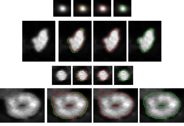
In oncology, Positron Emission Tomography (PET) imaging is widely used in diagnostics of cancer metastases, in monitoring of progress in course of the cancer treatment, and in planning radiotherapeutic interventions. Accurate and reproducible delineation of the tumor in the PET scans remains a difficult task, despite being crucial for delivering appropriate radiation dose, minimizing adverse side-effects of the therapy, and reliable evaluation of treatment. Our aim is to provide clinicians with intelligent software supporting accurate, efficient and reproducible delineation of the tumor.
Our Publication
Our Solution
- SterSEG – package for PET images segmentation
Our success
Automated segmentation of PET images for the delineation of tumor volumes has been the focus of intense research efforts for the last few years. There has also been a few limited efforts to compare several methods on common datasets, but in the majority of cases, each method has been evaluated on different image sets according to different evaluation criteria, and a comparison of currently available methods based on literature analysis only is thus challenging, if not impossible. A MICCAI challenge was an interesting opportunity to compare numerous existing methods implemented by their original authors (thus avoiding issues associated with re-implementation of methods by others) on a common set of images.
It’s good news: Our team won PET segmentation challenge using a data management and processing infrastructure!