RADIOLOGY Module
YOUR DICOM ANALYSIS BOOSTED
Radiology Module - faster and better treatment
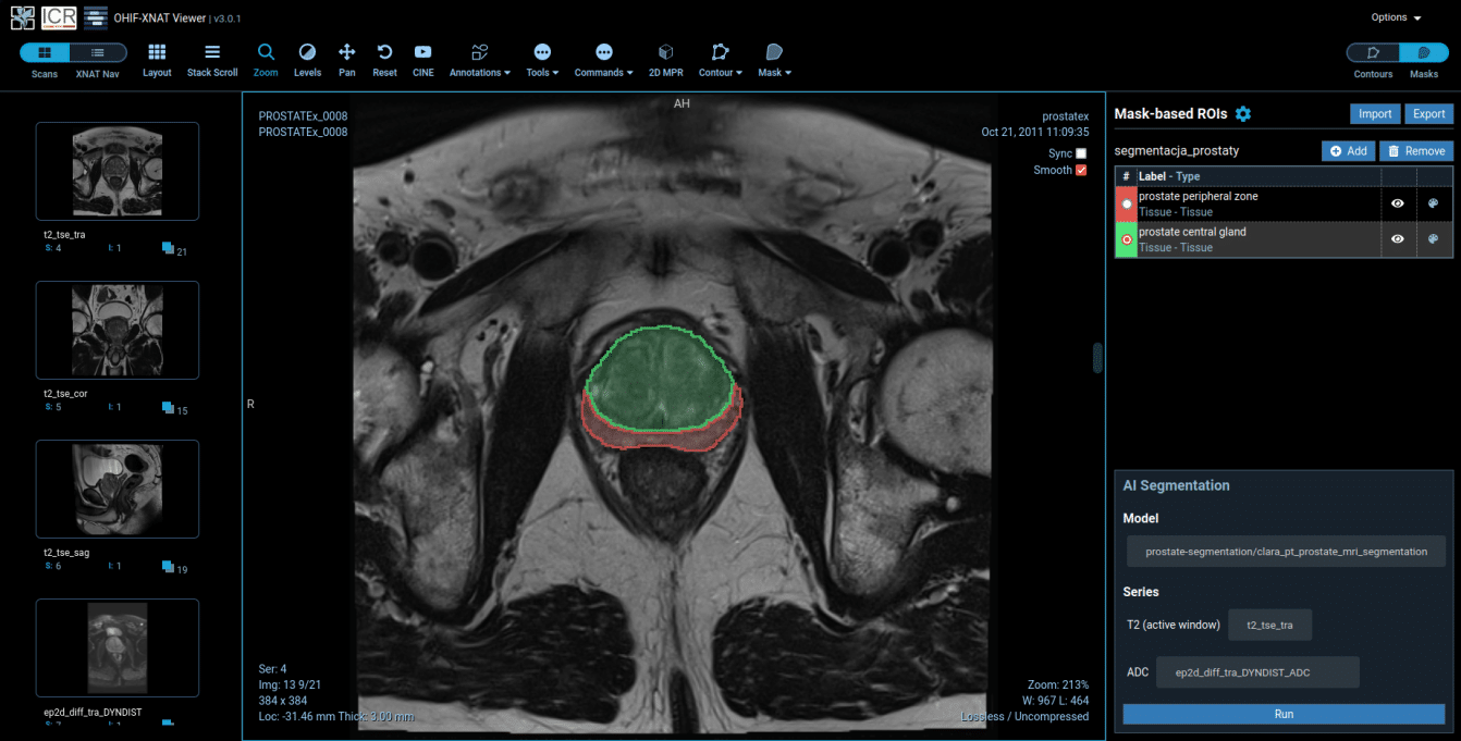
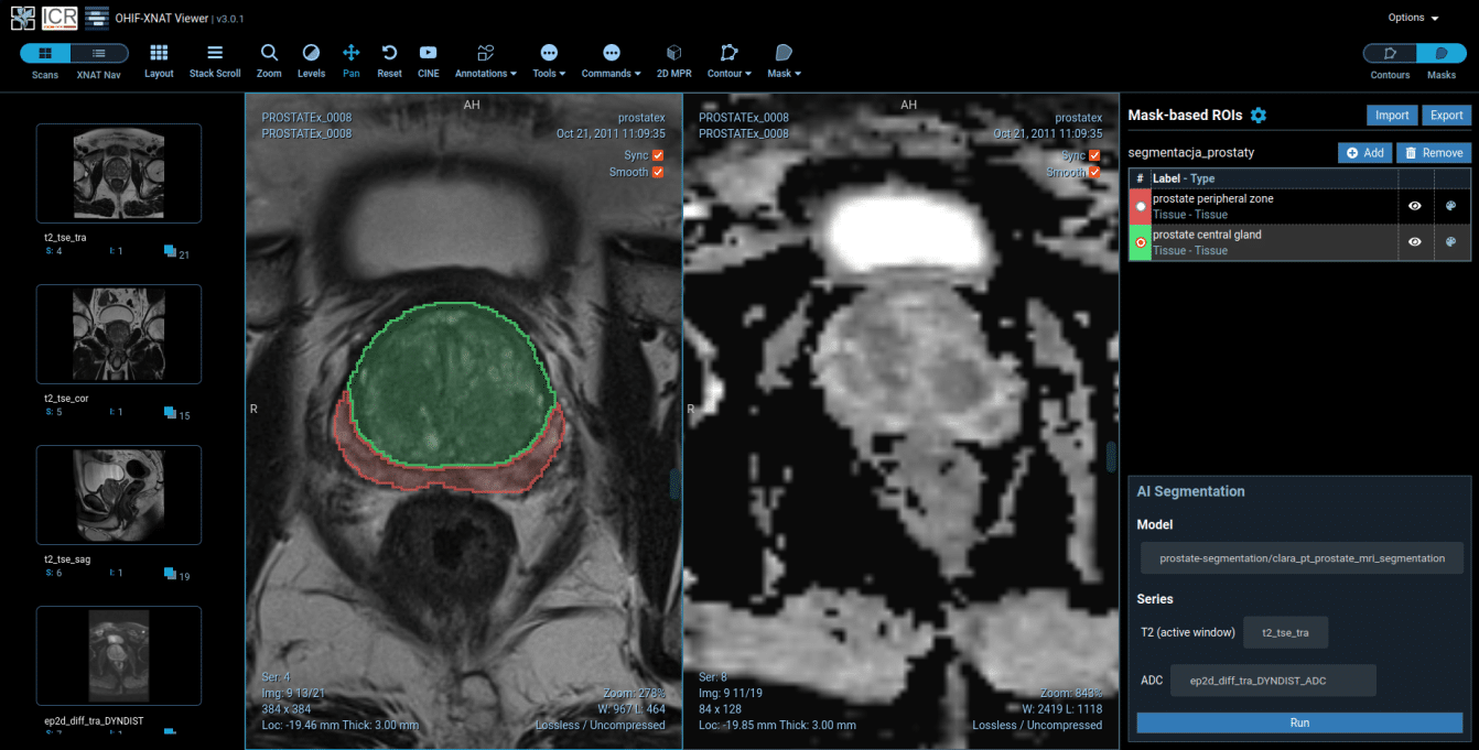
Radiology Module screenshots show an MRI scan of prostate (T2-weighted images in axial plane) that was segmented by our deep learning algorithm. The transitional zone is outlined in green and the peripheral zone in red.
Spend less time on single analyse and have more time to focus on truly challenging cases.
See how it works!
1. Firstly, acquire medical images
2. Secondly, store your data safely in the cloud
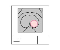
3. Next, use the software to find region of interests
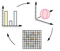
4. Finally, run algorithms for characterization
5. Use the power of deep learning
9. Faster and better treatment with Radiology Module
6. After that, double check your diagnosis & staging
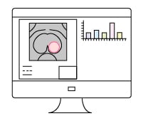
7. Collaborate with other experts from all over the world
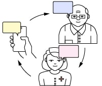
8. Lastly, generate reliable and accurate reports
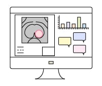
Greater productivity with AI
Plentiful, personalised, reproducible and objective dataPlentiful
Visual analysis can extract only about 10% of the information contained in a digital medical image. In contrary, Radiology Module converts the digital image into a huge quantity of data. To clarify, images are computed with specific mathematical algorithms. Extracted informations could not be processed by human eye in the same time. Moreover, the evaluated features describe imaging parameters as intensity, shape, size, volume, textures, etc. In other words, extracting an enormous wealth of quantitative data from medical images surpass humans possibilities.
Objective
In the first place, Radiology Module provides trained radiologists with pre-screened images and identified features. Not only data-driven approach allows for more abstract feature definitions but also makes it more informative and generalizable. Above all, diagnoses are more accurate thanks to built-in algorithms. In addittion, using algorithms reduce the risk of subjective description of tumour. Moreover, decision supported by AI can be shared with other specialists to resolve any doubts. To sum up, diagnoses are double checked, more precise and less subjective.
Personalized
As has been noted, Radiology Module can be used to investigate numerous characteristics. Similarly, tumours variability provides valuable information on both the aggressiveness and therapeutic response. Definitely, the more data extracted from different spatial and temporal levels, the more accurate diagnostic and prognostic models. Thanks to that, specialists can predict the right treatment, for the right patient, at the right time. Certainly, the future of patient care is dependent on artificial intelligence.
Reproducible
Radiology Module extracts qualitative and quantitative characteristics out of medical images. After that, experts provide details on size, location and morphology using built-in features. Meanwhile, complete confidentiality is ensured throughout the entire process. In other words, all extracted data are stored, viewed, shared and evaluated without data sharing and privacy concerns. Therefore, doctor to doctor cooperation within the organization as well as outside is highely improved.
We won MICCAI 2015 and 2016

Medical Image Computing and Computer Assisted Intervention 2015
Competitions in the field of “Imaging & Digital Pathology”
- 1st place in the competition in the Combined Radiology and Pathology Classification (Classification based on images of radiological and histological examinations)
- 3th place in Segmentation of Nuclei in Pathology Images (segmentation of cells in histological images).
Try Radiology Module for free
Run demo and benefit from the power of AI or contact us for more details.
