
KF-PRX slide scanners series
AI supported dependable partner for laboratories
KFBIO provides advanced digital pathology solutions recognized for their reliability, high-quality imaging, and laboratory workflow efficiency. The portfolio includes whole-slide scanners, slide printers, and other devices supporting the digitalization of pathology processes. As the official KFBIO distributor, Cancer Center delivers professional consulting, implementation, training, and technical support to ensure successful deployment and long-term performance.
From 20 to 1200 slides
KF-PRX series adapts effortlessly from routine tasks to large-scale digitization. Its dual-core high-speed engine and continuous loading enable true 24/7 continuous scanning, supporting all slide types with precision and consistency. Paired with an intelligent software suite, PRX streamlines storage, management, diagnostics, research, and multi-site collaboration.
True-to-Life Color, Built on Precision.
Our scanners deliver unmatched color fidelity through Independent Three-Channel Processing and the proprietary K-SCP (Specialized Color Processing) engine. This integrated system systematically enhances color, saturation, white balance, and contrast for exceptional image accuracy.
With built-in ICC profile calibration, colors remain consistent and reliable across all displays. From precise capture to faithful presentation, every digital slide is rendered perfectly trustworthy.
Unrivaled Optics, Unmatched Clarity.
We achieve a dramatic leap in optical resolution through holistic optimization of the entire imaging chain—from the light source and objectives to the camera and optical pathway. This premium configuration reveals exquisite cellular details, delivering images with razor-sharp nuclear edges and crisply defined internal architecture.
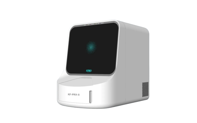
KF-PRX-S
Digital Pathology 20-120 Slides Scanner (PRX)
High throughput Digital Pathology Scanner.
Fast, Precise, Efficient,
Bright-field&Fluorescence,
Continous Loading, Clinic, Education, Remote Consultation
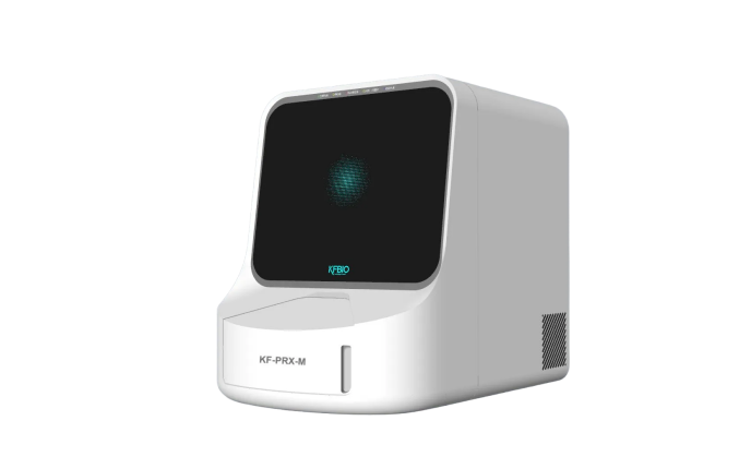
KF-PRX-M
Digital Pathology 150-480 Slides Scanner (PRX)
High throughput Digital Pathology Scanner.
Fast, Precise, Efficient,
Bright-field&Fluorescence,
Continous Loading, Clinic, Education, Remote Consultation
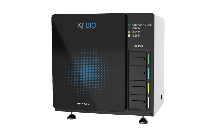
KF-PRX-L
Digital Pathology 640 Slides Scanner (PRX)
Ultra-high throughput Digital Pathology Scanner.
Fast, Precise, Efficient
Bright-field&Fluorescence,
Continous Loading, Clinic, Education, Remote Consultation
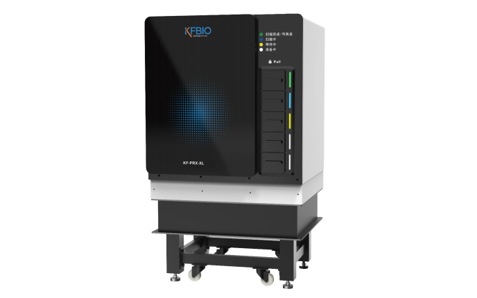
KF-PRX-XL
Digital Pathology 1200 Slides Scanner (PRX)
Ultra-high throughput Digital Pathology Scanner.
Fast, Precise, Efficient
Bright-field&Fluorescence,
Continous Loading, Clinic, Education, Remote Consultation
| IMAGING & CAPACITY | ||||
| Name | KF-PRX-S 20-120 | KF-PRX-M 150-480 | KF-PRX-L 640 | KF-PRX-XL 1200 |
| Brightfield | ||||
| Objective Type | 20x 0.8NA & 40x 0.95NA | |||
| Optical Magnification | 20x & 40x | |||
| Speed | 15 sec / slide , 20x 25 sec / slide, 40x | |||
| Throughput | up to 90 slides / hour | up to 90 slides / hour | up to 100 slides / hour | up to 200 slides / hour |
| Scan Core Numbers | 1 | 1 | 1 | 2 |
| Resolution | 0.36 um/pixel at 20x, 0.18 um / pixel at 40x | |||
| Scan Method | Line Scanning | |||
| Output Format | .KFB, .SVS, .DCM, .TIFF | |||
| Fluorescence | ||||
| Objective Type | 20x 0.8NA & 40x 0.95NA | |||
| Optical Magnification | 40x & 80x | |||
| Speed | ≤6 min / slide, 10mm * 10mm, 3 filters ≤12 min / slide, 10mm * 10mm, 6 filters | |||
| Scan Core Numbers | 1 | 1 | 1 | 1 |
| Resolution | 0.33 um/pixel at 20x, 0.17 um / pixel at 40x | |||
| Scan Method | Area Scanning | |||
| Output Format | .KFBF, .QPTIFF | |||
| Upgrade to Support Fluorescence | Yes | Yes | Yes | Yes |
| Fluorescence Channel Options | 3,4,6,8,10 | 3,4,6,8,10 | 3,4,6,8,10 | 3,4,6,8,10 |
| Common Paramters for Both Brightfield and Fluorescence | ||||
| Field of View (FOV) | 1mm | |||
| Z-Stack Scanning | ≤10 layers | |||
| Multi Layer Image Fusion | Support | |||
| Objective Lens | Evident X-Line UPLXAPO 20X, UPLXAPO 40X, Optional 4X / 10X / 50X / 60X | |||
| Objective Lens Sockets | 5 | |||
| Slide Capacity | 20~120 | 150~480 | 640 | 1200 |
| Slide Holder Type | Rack | Rack | Rack | Rack |
| Slide Holder Capacity | 20 | 20 or 30 | 20 | 20 |
| Slide Holder Number | Maximum 4 racks | Maximum 16 racks | 32 | 60 |
| Direct Placement of Racks With Handles Intact | Yes | Yes | No | No |
| Capacity for the Rack with Handle | Maximum 60 slides, 3 racks * 20 slides | Maximum 240 slides, 8 racks * 30 slides | – | – |
| Slide Pre-loading | No | No | Yes | Yes |
| Continuous Loading | Support | |||
| Slide Order Sorting Support | No | No | Yes | Yes |
| Label Identification | Support | |||
| DIMENSIONS & WEIGHT & POWER | ||||
| Scanner Dimensions (W x L x H) | / | 740*530*730 | 645*820*790 | 800*890*1535 |
| Power Supply Voltage Range | 100–240V AC, 50/60Hz | |||
| Power Socket Type | Type A、Type C、 Type D、Type E/F、Type G、Type I、Type J、Type L、Type M | |||
| IT & COMPUTER | ||||
| PC Model | Dell OptiPlex 7020MT Plus | |||
| PC Parameter | CPU: I5-14500 Memory: 32G*2 Harddisk: 1T SSD NVME | |||
| Monitor | 24 Inch | |||
| Network Adopter | 1Gb RJ45 * 1 | |||
| Operation System | Windows 10 Professional 64bit | |||
| Slides | ||||
| General | The tissue/smear must be mounted with a coverslip, glass or tape. | |||
| Standard | 1-inch x 3-inch (2.54 cm x 7.62 cm) slides. Compatiable with ISO 8037/1. | |||
| Thickness | 0.9 mm ~ 1.1 mm, excluding coverslip | |||
| Special Requirement | The coverslip and attached label must remain fully within the microscope slide perimeter, secured with complete adhesive contact. All surfaces shall lie flat without edge separation. The slide exterior must maintain dryness with no fluid leakage from the specimen zone. | |||
| Maximum Tissue/Smear Thickness | Optimized for tissue thickness of 3-5 μm (including mounting media) | |||
| Coverslips Accepted | The coverslip/label assembly (0.17 mm nominal thickness, standard borosilicate glass or cellulose tri-acetate film) must maintain full edge registration with the microscope slide substrate through continuous adhesive bonding. All interfacial surfaces shall exhibit co-planar adhesion integrity with zero delamination, while the external slide surfaces remain contamination-free from residual fluids or specimen migration. | |||
| Label Area (W x L) | 25 mm x 25 mm | |||
| Label Special Requirement | Adhesive labels must maintain non-protrusive margins within slide boundaries and demonstrate full-surface lamination to the coverslip interface plane (upper slide surface). Prohibited from substrate attachment on the specimen observation surface (lower slide plane), all labeling components shall exhibit edge-to-edge planar adhesion without delamination defects. | |||
| Barcodes Supported | 1D and 2D Barcode, QRCode, DataMatrix Code | |||