MRI Analysis

Our Mission
Our mission is to combine state of the art computer science research with medical imaging to improve the quality of diagnosis, save doctor’s time and ultimately to give patients a better chance. We hope that one day with the help of our solutions doctor’s would have more time to focus on truly challenging cases, that they would have the tool to double check their diagnosis in case of any doubt and that patients would be able to commence the appropriate treatment sooner.
The Solution
We believe that in order to make a world of cancer treatment a better place we need to make use of the technology to the fullest. We think that using advanced machine learning techniques in medical image diagnosis on the regular basis is the next big leap that our society needs to make in order to progress and we are more than willing to facilitate this transformation.
See also web dicom viewer (web radiology tool): https://radiology.cancercenter.eu and our presentation.
Our case study
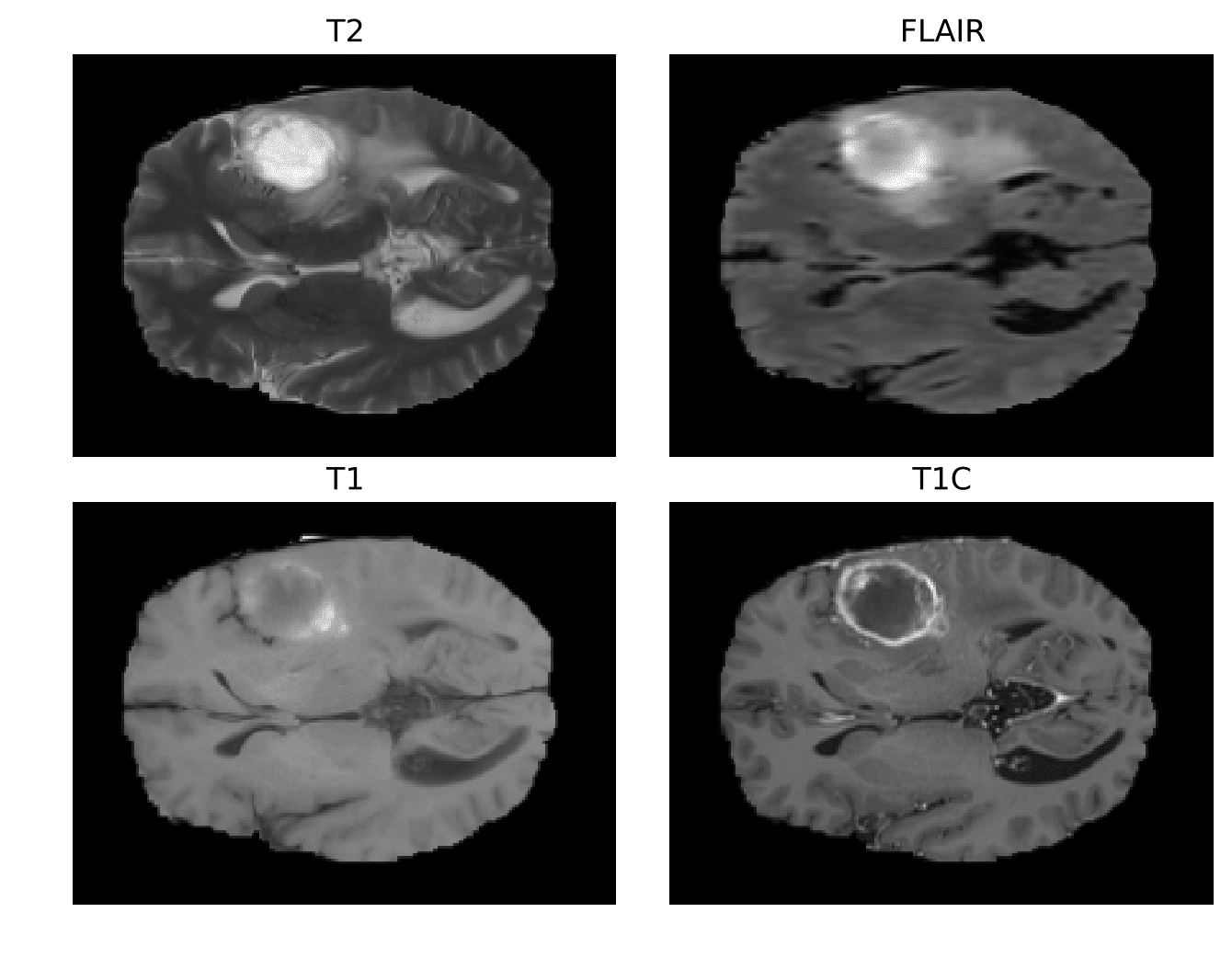
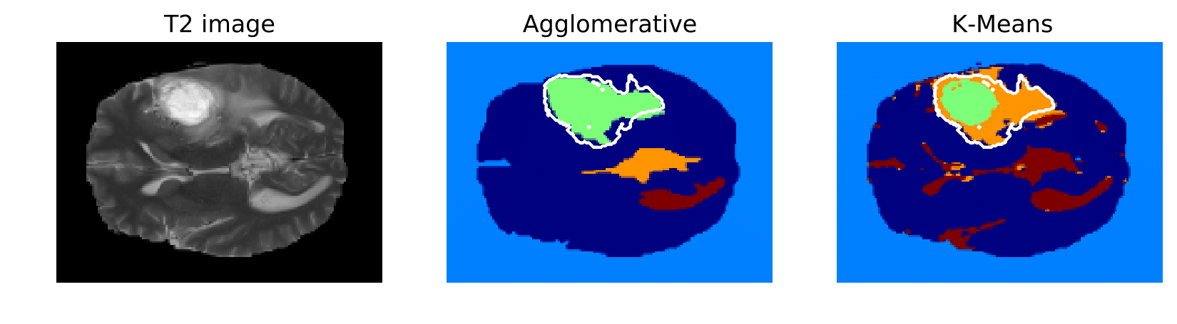
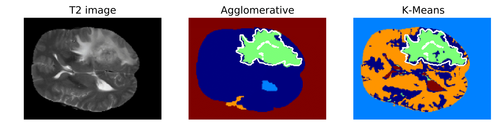
Radiology images of the same patient varied in terms of shape, average intensity level and head position. Both Otsu and adaptive method based on the histogram peaks (see Section 2.3) were able to extract binary mask of the head. The challenging part was to select the proper cluster, that corresponds to the tumor area. We were unable to do that using only single (primary) segmentation algorithm. Symmetry analysis of the hemispheres proven to be versatile tool for the cluster classification (whether it is a tumor or not). In the areas that was not affected by tumor, the asymmetrical differences were subtle.
Intensity, shape, position and textural features were extracted from the T1, T1C, FLAIR and T2 scans. Based on evolution and selection of features tumor position and distribution of intensity in FLAIR image was most relevant. According to the literature Oligodendroglioma often occurs in frontal and temporal lobes, it proven to be truth in my data set. If we would have only the position of the tumor, cross-validation classifier would have an accuracy of 70% (std: 12). Distribution of intensity in FLAIR scan had high correlation and the highest importance. In the T2 scans lesions always had high intensity, however in FLAIR lesion were either partially bright or dark. Textural pattern based classification requires more samples to be effective. Random Forest classifier validated with k-fold cross validation had average accuracy of 87.0% (std: 12.991).